


With the osteochondrosis of the spine, many are not familiar not to the popular gears of the TV screen, but on their own sad experience.The statistics are severe: up to 80% of the population suffers from this disease, which also much younger.If the previous complaints concerning problems in the spine were mainly older generation, children 'osteochondosis no longer surprises anyone.And the fault of a sedentary lifestyle and the "advantages of civilization".

Osteochondrosis of the cervical column is a progressive polietiological disease which manifests itself by the degeneration of intervertebral discs and the dystrophy of the ligament apparatus of the spine.Everyone knows first -hand symptoms, but this knowledge is fragmentary;We will try to structure them, as well as to talk about the principles of diagnosis and the treatment of osteochondrosis of the cervical column.
The causes of osteochondrosis
Medical science cannot respond unequivocally, which is why osteochondrosis occurs.It is reliably known that the sedentary lifestyle that a modern person is subject to negatively affects the progression of this disease.It is interesting to note that hypodynamia and colossal loads of athletes lead to a proxy of discs.A hereditary factor plays a leading role.The following reasons are distinguished:
- Load hereditary history;
- obesity;
- Hypodynamia;
- metabolic disorders in the body;
- traumatic damage to the spine;
- Long static overloads and work associated with lifting weights (work on computer, weight lifting, minors, movers, etc.);
- Scoliosis;
- dysfunctional environmental situation;
- flat feet and pregnancy;
- Hypothermia and stress, which often cause exacerbations of the disease.
There are several neurological syndromes:
- Speed-shoulder periarthritis;
- root;
- heart;
- Vail artery syndrome.
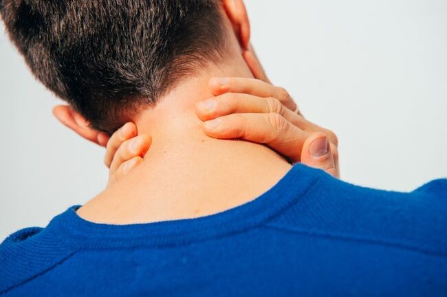
Speed-shoulder periarthritis.It is characterized by pain in the neck, shoulder, shoulder joint.The main neurogenic contracture of the shoulder joint is formed, which is of a protective nature, because it protects the axillary nerve against stretching (analgesic installation).With this position, the muscles surrounding the joint are in tension.The severity of pain syndrome depends on the degree of exacerbation of osteochondrosis: a slight limitation of the amplitude of movements in the "frozen shoulder" articulation if called, when movements cause serious pain.The pain intensifies when the shoulder is diverted and pronounced, because it is these movements that improve the tension of the axillary nerve.
Roshift syndrome (Cervical Radiculitis).Most often, it occurs with cervical osteochondosis.At the same time, the spine of the nerve of the spine is pressed due to the "sagging" of the intervertebral discs, as well as because of the growth of osteofites or the projection of the discs in the lateral direction.Pain syndrome is specific: intense burn, tear and pressing, which also intensifies when the patient moves his head.The analgesic installation is also noted in the neck muscles, they are strongly tense and painful, the volume of movements is limited.There is pain on the back of the head, neck, front chest, shoulder, between the shoulder blades.The disruption of sensitivity by the type of "half jackets with short sleeves" is characteristic.
Cardial syndrome.The name of the syndrome is responsible for itself: the clinical image is very similar to angina.In this case, there is no organic damage to the heart, at the height of pain syndrome, violations of coronary blood flow by ECG are not detected and these patients are well tolerated.A typical characteristic with angina: the pain takes place after taking nitrates and, in the case of osteochondosis, does not change and disturbs for a long time.Unlike angina, the location of pain is mainly in the heart on the left.With the irritation of the roots of the C8 - T1 segments, rhythmic disturbances in the form of tachycardia and extrasstole are possible.This is not due to damage to the driver's system of the heart, but to a violation of the sympathetic innervation of the heart muscle (extracardian damage).In the differential diagnosis of angina of chest and cardiac syndrome, the leader is the fact that, in addition to heart complaints, the patient notes the increase in pain in the shoulder joint and the neck associated with lifting or severe movements.
Vail artery syndrome.The vertebral artery takes place in a channel formed by holes in the transverse processes of the vertebrae.This artery is twinned, it is responsible for the blood supply to the brain.Consequently, any narrowing of this channel very negatively affects the nutrition of brain tissues.The vertebral artery syndrome develops directly both with the compression of the artery itself and with the irritation of the sympathetic nervous plexus, which is located around it.The pain in this pathology is burning or pulsating in the occipital region with a propagation to whiskey, tutorial arches, crows.It arises on both sides.Patients generally associate worsening with disease after sleep in non -physiological installation, transport trips, a walk.With pronounced symptoms, hearing loss, stunning, noise in the ears, nausea, vomiting, loss of consciousness and increasing blood pressure are possible.These symptoms are not specific and are very similar to the complaints of the brains.This pathology is characterized by the Sistine Chapel syndrome: a fainting that occurs when you overturn your head back (severe cerebral ischemia).It was described by visitors to the Vatican sixtine chapel when they examined the frescoes of its arches.It is also possible to fall without loss of consciousness with clear turns of the head.
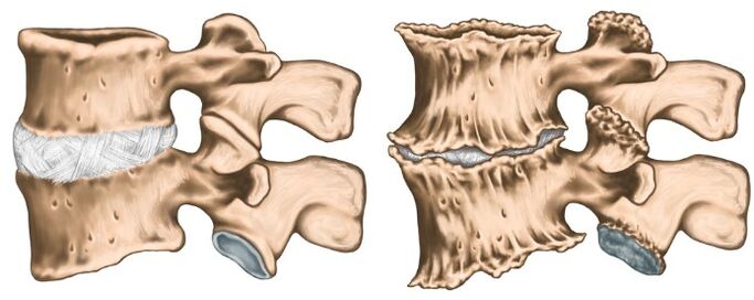
Like any diagnosis in medicine, the diagnosis of osteochondrosis is established on the basis of patient complaints, the anamnesis of the disease, the clinical examination and the auxiliary research methods.The X -ray of the cervical column in direct and lateral projections is carried out, if necessary in special positions (with an open mouth).At the same time, experts are interested in the height of the intervertebral discs, the presence of osteofites.Modern research methods, IAMR and CT research is used, which makes it possible to check the diagnosis most precisely.In addition to the methods listed of additional research, consultations with related specialists (cardiologist, ophthalmologist, neurosurgeon) may be necessary and the examination of the neurologist is simply vital.The neurologist is engaged in the treatment of osteochondrosis, so after examining the patient, he will prescribe the minimum examination necessary for his discretion.
Treatment of osteochondrosis
Osteochondrosis is a polyetiological disease because a therapy lesson is not cured.You cannot drink a "magic pill" and everything will pass, it is necessary to fundamentally change your lifestyle, because the trigger is hypodynamia.The most tangible results are easier to achieve at the initial stage of the disease, when complaints are minimal and there are no compression syndromes and the vertebral artery.In the acute stage of the disease, when the following groups of drugs are prescribed in pronounced pain: pain syndrome is pronounced:
- Therapeutic paravertebral blocking (to relieve pain and the elimination of muscle spasms);
- NSAIDs;
- nails containing NSAIDs and a reflex action;
- muscle relaxing;
- B Vitamins V.
As the inflammatory process lights up and the relief of pain syndrome, they switch to the treatment of physiotherapy.Most often, the following techniques are used:
- Laser therapy;
- electrophoresis;
- acupuncture;
- Exercise therapy;
- Therapeutic massage;
- Manual therapy.
It is important to understand that osteochondrosis proceeds to periods of exacerbation and remission, it is therefore very important to affect the cause and not to deal with the investigation.