


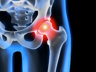
Arthrosis of the hip joint — degenerative-degenerative pathology, which is characterized by destruction of hyaline cartilage. The disease develops gradually, together pain syndrome and decreased range of motion. In the absence of medical intervention at the initial stage of arthrosis several years later occurs atrophy of the thigh muscles. The injured limb is shortened, and the fusion of the articular cleft leads to partial or complete paralysis of the hip joint. The causes of the pathology are of a previous injury, curvature of the spine, systemic disease of the musculoskeletal system.
Osteoarthritis usually detected in patients of middle age and older people. Diagnosis is established on the basis of the results of instrumental research — x-ray, MRI, CT, arthroscopy. Treatment pathology 1 and 2, the degree of severity of a conservative. When findings of ankylosis or ineffectiveness of medical therapy is performed surgery (arthrodesis, arthroplasty).
The mechanism of development of pathology
In the hip joint consists of two bones — the iliac and femoral. The lower separation of the iliac bones represented her body, which deals with the plexus with the femur, forming the upper department of the acetabulum of the trough. During the movement of the articular fossa immobilized, and the femoral head moves freely. It's the "swivel" device hip the completion of the joints allows him to flex, stretch, rotate, supports abstraction, submission of the thigh. The smooth sliding of the articular structures provides a smooth, elastic, flexible hyaline cartilage lining the cavity, and the head of the femur. Its main function is the redistribution of load during movement, notice the rapid fraying of the bone tissue.
Under the influence of external or internal factors is violated trofika cartilage. Does not have its own circulatory system — the nutrients it supplies the tissue of the synovial liquid. When osteoarthritis it thickens, it becomes more viscous. Discovered a lack of nutrients causes a drying out of the surface of the hyaline cartilage. She is covered with cracks, which leads to a continuous micro-injuries to tissues during flexion or extension of the hip joint. Cartilage narrowing, losing its depreciation characteristics. To "adapt" to the increase in pressure, deformed bones. And on the background of the deterioration of metabolism in the tissues of the progress is destructive-degenerative changes.
Causes and precipitating factors
Idiopathic or primary osteoarthritis develops without any reasons. It is believed that the destruction of the cartilage tissue occurs because of natural aging of the organism, slows down the regenerative processes, reduce the production of collagen and other substances, necessary for the full regeneration of the structures of the hip joint. Secondary osteoarthritis arises on the background of already present in the body of the defective condition. The most frequent reasons of secondary disease include:
- previous injury — injury to the tendon apparatus, a break in the work of the muscles, their complete detachment from the bone reason, fractures, sprains;
- violations of the completion of the joints, congenital dysplastic disorders;
- autoimmune disease — rheumatoid, reactive, psoriatic arthritis, systemic lupus erythematosus;
- non-specific inflammatory diseases, for example, the festering arthritis;
- specific infections — gonorrhea, syphilis, brucellosis, ureaplasmosis, trichomoniasis, tuberculosis, osteomyelitis, encephalitis;
- disruption of the functioning of the endocrine system;
- degenerative-degenerative pathology — osteochondropathy head of the femur;
- hypermobility of the joints, drive the production of "over strength" of collagen, provoking their excessive mobility, weakness of the ligaments.
So as a cause of the development of arthrosis can become a hemarthrosis (bleeding into the cavity of the hip joint), for which causing factors include blood disorders. Prerequisites to the emergence of diseases are the extra weight, excessive physical load, a sedentary way of life. To its development lead to the incorrect organisation, sports training, lack in the diet of foods with a high content of trace elements, Giraud - and water-soluble vitamins. Postoperative arthrosis arises a few years later after surgery, especially if it is accompanied by excision of a large volume of tissue. Arthrosis of the hip joint can not be transmitted by inheritance. But in the presence of certain congenital features (disorder of the metabolism, the construction of the skeleton) the likelihood that its development is significantly increased.
Symptoms
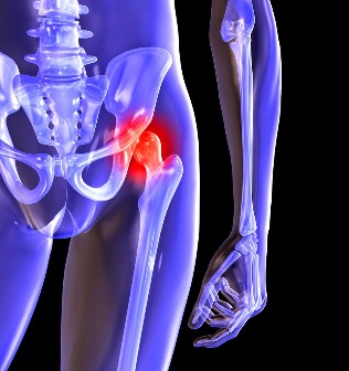
The head of the symptoms of the arthrosis of the hip joint — pain when walking in the area of the thighs, the knees of an articulated arm. A person suffering from stiffness of movements, rigidity, and especially in the morning. To stabilize the joint, the patient begins to limp, it changes his walk. With time due to muscle atrophy and deformity of the completion of the joints of the limb considerably shortened. Another characteristic of the pathology — the restriction area of the thigh. For example, difficulties arise when trying to sit down on a chair, with his legs apart on the side.
Even the "running" OSTEOARTHRITIS can be treated at home! Just remember to once a day primer this...
For arthrosis of the first degrees of severity are characterized by recurrent pain incurred after intense physical exertion. They localized in the area of the completion of the joints and disappear after a longer vacation.
In osteoarthritis the second degree of the hip links the severity of the pain syndrome increases. Uncomfortable feelings arise even at rest, apply to the thighs and groin, are amplified when lifting weights, or increasing physical activity. How to remove pain in the hip joint, the person begins to barely noticeable limp. Celebrate the restriction of movements in the joint, and especially in lead and internal rotation of the hip.
Osteoarthritis in the third degree is characterized by permanent severe pain. During the movement of the problems arise, so when walking one is forced to use a cane or crutches. Because of the weakness, which emit thigh muscles occurs displacement of the pelvic bones in the frontal plane. To compensate there has been a shortening of the legs of the patient when the movement leans to the side of the damaged limb. It induces a strong shift of the center of gravity and increase the load on the articulated arm. At this stage of the arthrosis develops significant stiffness of the joint.
| Extent | Radiographic signs of |
| The first | Changes expressed not dramatically. Joint space slightly and irregularly constricted, lacking to the destruction of the surface of the femur. On the outer or inner edge of the acetabulum depression observed small bone growths |
| The second | The height of the articular slit is significantly reduced as a result of its uneven seams. Bone crown modified thigh up, deformed, enhanced, its contour becomes uneven. Bone growths arise on the surface of the inner and outer edges of the articular fossa |
| The third | There was a complete or partial fusion of the articular cleft. The head of the femur is strongly expanded. More bone growths located on the surface of all the acetabulum depression |
Diagnosis
When placing a diagnosis the doctor takes into account the clinical manifestations of the pathology, medical history, results of external examination of the patient and instrumental studies. The most informative radiography. With its help it is assessed the condition of the hip joint, is set to the degree of its swing, the degree of damage to the cartilage tissue, and in some cases the reason for the development. If the cervical-diaphyseal node increased, and vertluzhnoj depression beveled and flattened, what with the big degree of probability it can be assumed dysplastic congenital changes of the completion of the joints. The disease Pertusa points out the broken shape of the femur. Radiography allows vyvit post-traumatic osteoarthritis, despite the absence of a history of previous infection injuries. Also, use other diagnostic methods:
- CT helps us to discover the growths the edges of the bone plates, these body parts of the osteophytes;
- MRI is done to assess the condition of the connective tissue structures and the extent of their involvement in the disease process.
If necessary, the inner surface of the completion of the joints examined using arthroscopic instruments. Differential diagnosis is performed for the exclusion of gonarthrosis, the lumbar-sacral or thoracic degenerative disc disease. Pain in osteoarthritis may issue for the clinical manifestations of radicular syndrome, caused by violation or inflammation of the nerve. To exclude neurogenic pathology usually succeed with the help of several tests. Arthrosis of the hip joint necessarily changing from the top of bursitis of the hip joint disease ankylosing Spondylitis, reactive arthritis. For exclusion of autoimmune diseases are biochemical examination of blood and synovial fluid.
Surgery
When ineffectiveness of conservative therapy il diagnosis of complicated pathology is carried out the operation. To restore the cartilage tissue in the joint, the damaged joint with arthrosis of suffering, without surgery, prosthetics impossible, but with the right approach to treatment, compliance with all medical regulations, governed by the proper lifestyle, the persecution of the therapeutic gymnastics, regular classes massages, taking vitamins and proper nutrition it is possible to stop the process of destruction and the destruction of cartilage and hip joints.